Muscles Of The Floor Of The Mouth Ct
Http Pdf Posterng Netkey At Download Index Php Module Get Pdf By Id Poster Id 110081

Floor Of Mouth Anatomy Radiology Coronal Google Search
Http Pdf Posterng Netkey At Download Index Php Module Get Pdf By Id Poster Id 101807

Pin By Dr Abuaiad On Brain Head And Neck Head And Neck Radiology Image

Orbital Rhabdomyosarcoma Soft Tissue Mass In The Inferior Aspect Of The Orbit Inseparable From Inferior Rectus Muscle The Mass Results In Proptosis The Globe

Pin By Dr Abuaiad On Brain Head And Neck Molar Tooth Lymph Nodes Head And Neck
The floor of mouth is an oral cavity subsite and is a common location of oral cavity squamous cell carcinoma.
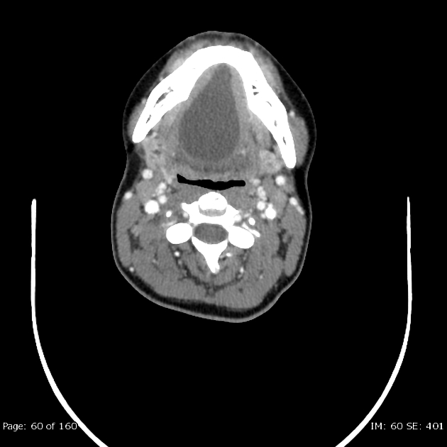
Muscles of the floor of the mouth ct. The muscles of the mouth human anatomy. The mylohyoid genioglossus and geniohyoid muscles comprise the muscular floor of the oral cavity. The plane is medial to the mylohyoid m muscle and contains the hyoglossus h muscle. Oral cavity and floor of the mouth.
Mancuso and robert hermans imaging approach techniques and relevant aspects general examination patients should be positioned with the neck extended so that the mandibular body is perpendicular to the tabletop for either computed tomography ct or magnetic resonance mr study. Muscles of floor mouth ct anatomy. Whats people lookup in this blog. Coronal ct a and t1 weighted mr b images at the middle of the floor of the mouth show the sublingual space as a plane with low attenuation at ct and high signal intensity at mr imaging.
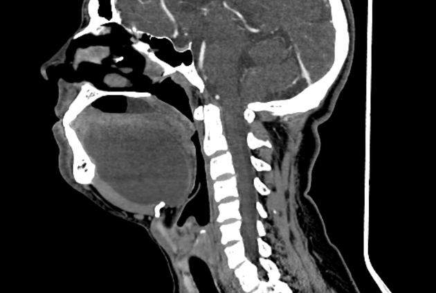
The floor of mouth is bounded anteriorly and laterally by the lower gingiva medially by the oral tongue and posteriorly at the insertion of the anterior tonsillar pillar into the oral tongue. Its medial fibers form the angular head which arises by a pointed extremity from the upper part of the frontal process of the maxilla and passing obliquely downward and lateralward divides into two slips. The quadratus labii superioris is a broad sheet the origin of which extends from the side of the nose to the zygomatic bone.

Pin On Excalibur S Mri Board

Axial Ct Section Of The Floor Of The Mouth Showed A 1 38 1 18 In Download Scientific Diagram
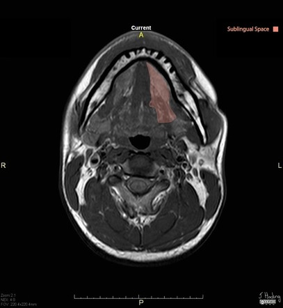
Sublingual Space Radiology Reference Article Radiopaedia Org

Pin By Dr Abuaiad On Brain Head And Neck Darth Head And Neck Darth Vader

Floor Of Mouth The Term Floor Of The Mouth Is Used Differently By Different Authors But In All Cases It Is Ap In 2020 Muscle Anatomy Facial Nerve Medical Illustration
Floor Of Mouth Dermoid Cyst Radiology Case Radiopaedia Org

Coronal Head Neck

Epos

Image Result For Jugulodigastric Lymph Node Location Lymph Nodes Nasal Cavity Cervical

Oral Cavity And Floor Of The Mouth Introduction Radiology Key

Review Of Imaging Anatomy And Pathology Of The Floor Of The Mouth Semantic Scholar
Floor Of The Mouth Cystic Lesion Radiology Case Radiopaedia Org
Https Pubs Rsna Org Doi Pdf 10 1148 Rg 315105062

