Orbital Floor Fracture On Panoramic Xray

An Orbital Blow Out Fracture Occurs When There Is A Fracture Of One Of The Walls Of Orbit But The Orbital Rim Remains Intact T Radiology Sinusitis Radiography
Traumatic Orbital And Occular Injury Radiology Key

Black Eyebrow Sign Black Eyebrow Sign Is The Description Given On Plain Facial Radiographs To Intra Orbital Air Air Rises Into Radiology Air Rise Air Leaks

Orbital Blowout Fracture Teardrop Sign Blowout Herniated Ocular
The Facial Bones

Orbital Floor Fractures Ento Key

Bilateral frontal intraparenchymal hemorrhages.
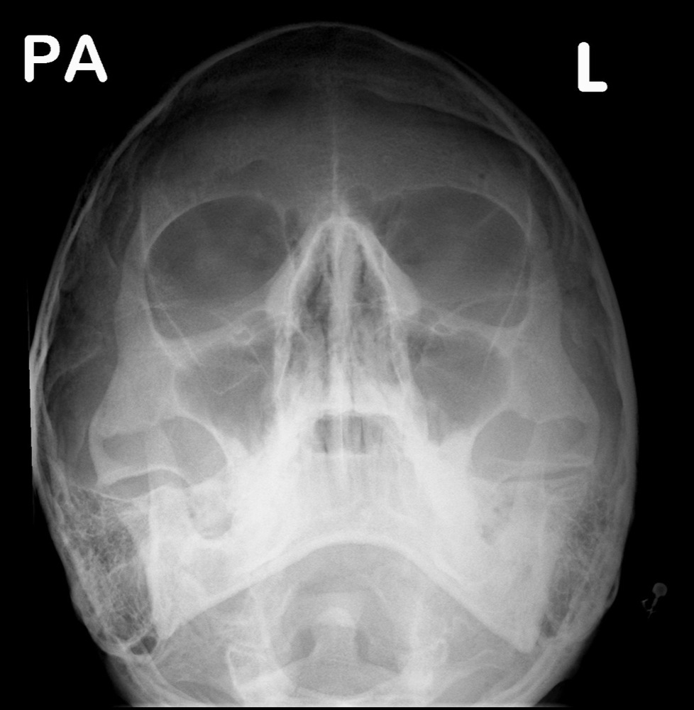
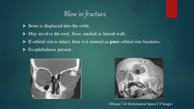
Orbital floor fracture on panoramic xray. 11 ultrasound is sometimes used for visualisation of the zygomatic arch and anterior wall of the frontal sinus particularly following reduction to avoid further radiation exposure. There are three types of orbital fractures. No evidence of rectus muscle entrapment retrobulbar hemorrhage or proptosis. If the patient is upright when the film is taken an air fluid level can often be seen in the maxillary sinus which may indicate fracture of the maxillary sinus orbital floor.
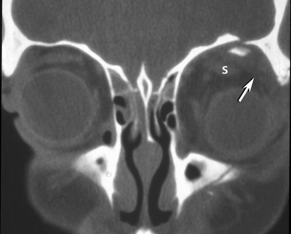
Getting hit with a baseball or a fist often causes a orbital blowout fracture. A crack in the very thin bone that makes up these walls can pinch muscles and other structures around the eye keeping the eyeball from moving properly. Getting hit with a baseball or a fist often causes a blowout fracture. Displaced malar complex fractures may increase orbital volume due to angulation of the zygomaticosphenoid suture or orbital floor blowout.
A blowout fracture is a break in the floor or inner wall of the orbit or eye socket. X ray and computed tomography scans may also be taken. Blowout fracture a break of the thin inner wall or floor of the eye socket. Getting hit with a baseball.
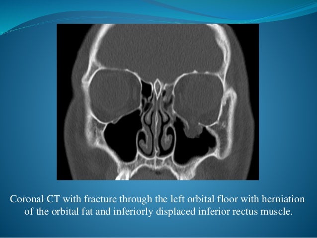
The blowout fracture is the most common type of orbital fracture and is usually the result of trauma. Hemorrhage partially fills the left maxillary sinus. This is reflected in the demographics. Orbital floor fractures may result when a blunt object which is of equal or greater diameter than the orbital aperture strikes the eye or on the cheek 1.
It is more prevalent in young men. Left orbital floor fracture. Orbital floor fracture also known as blowout fracture of the orbit eye socket. Waters view best displays inferior orbital rims nasoethmoidal bones and maxillary sinuses.

Black Eyebrow Sign Black Eyebrow Sign Is The Description Given On Plain Facial Radiographs To Intra Orbital Air Air Rises Radiology Black Eyebrows Air Rise

Facial Fractures Radiology Reference Article Radiopaedia Org

Orbital Blowout Fracture Wikipedia The Free Encyclopedia Blowout Fracture Nuclear Medicine

Orbital Blowout Fracture Radiology Reference Article Radiopaedia Org
Orbital Floor Blow Out Fractures

Pin On Ent Rotation

Pin By Kapil Moodley On Cranio Blowout Fracture Radiologic Technology

Femoral Neck Stress Fracture Radiology Case Radiopaedia Org Radiology Neck Fracture Emergency Medicine

Pin On Ct Scans
Orbital Fracture

Imaging The Face Radiology Key

Right Upper Lobe Pneumonia Radiology Case Radiopaedia Org Radiology Radiology Imaging Pneumonia

Facial Trauma Undergraduate Diagnostic Imaging Fundamentals

