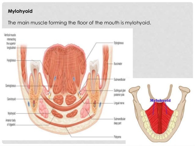
Muscle Forming Floor Of Oral Cavity
Oral Cavity Pharynx Radio Anatomy

Oral Cavity Anatomy Sagittal View

Oral Cavity Ppt Video Online Download

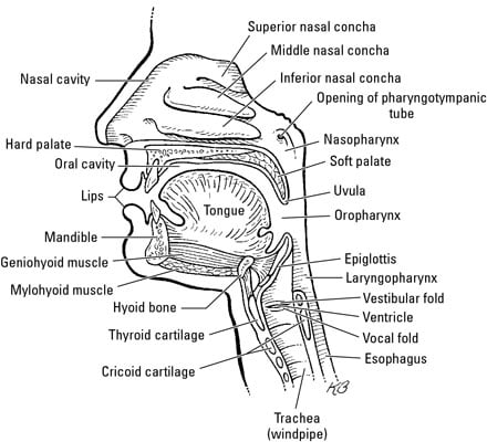
Anatomy Of Oral Cavity Pharynx And Esophagus

An3 08 Oral Cavity Oropharynx Swallowing At Touro University Nv Studyblue

The Oral Cavity Comparative Oral Ent Biology Openstax Cnx
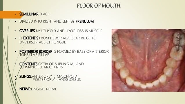
Muscles that help form the floor of the oral cavity.
Muscle forming floor of oral cavity. If a person has bad oral hygiene a cavity can form in the space between the crown and the real tooth. It is bounded by a roof a floor and lateral walls. Structures that pass through the aperture includes muscles. The floor of mouth is an oral cavity subsite and is a common location of oral cavity squamous cell carcinoma.
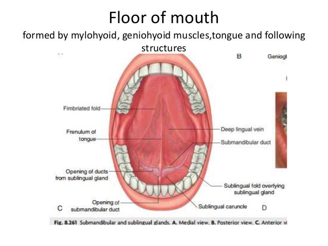
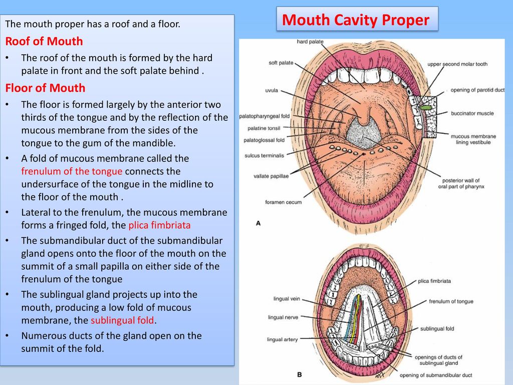
It is bordered by the hard palate in the upper front while the soft palate constitutes the upper back boundary 10 inferiorly it has various oral muscles glands tongue attachment to the oral cavity floor 7 through the flexible band of tissues known as lingual frenulum 21. The mylohyoid muscle is derived from the first pharyngeal arch. These muscles are mesodermal in embryologic origin. The floor of mouth is a u shaped space which extends and includes from the oral cavity mucosa superiorly and the mylohyoid muscle sling 2 3.
A cavity can form under a crown. Anteriorly it opens to the face through the oral fissure while posteriorly the oral cavity communicates with the oropharynx through a narrow passage called the oropharyngeal isthmus also termed the. It is named after its two attachments near the molar teeth mylo comes from the greek word for molar. The floor of mouth is bounded anteriorly and laterally by the lower gingiva medially by the oral tongue and posteriorly at the insertion of the anterior tonsillar pillar into the oral tongue.
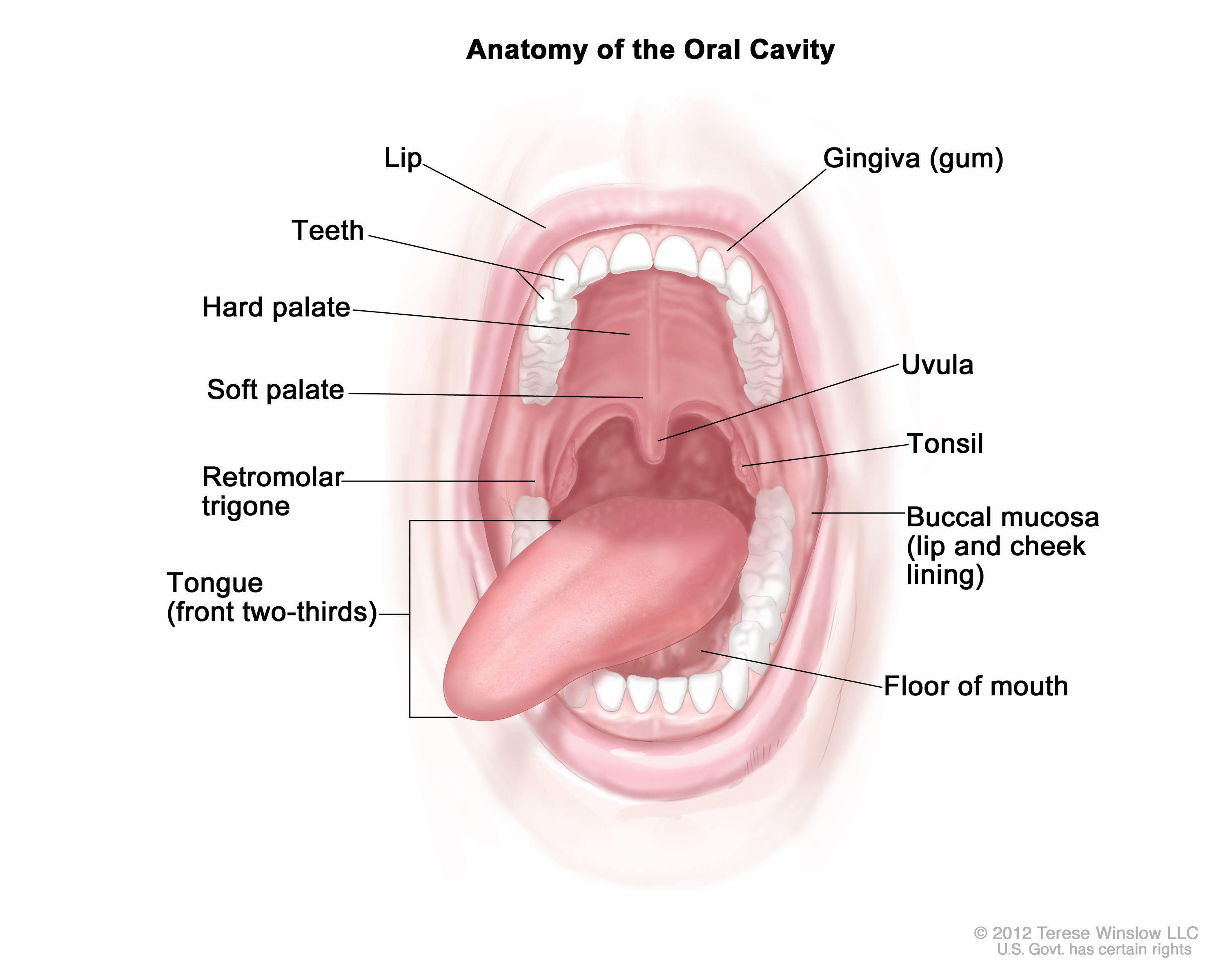
The posterior part of the oral cavity is referred to as the mouth proper. If a crown is not well adapted it can gather up dental. The free posterior border of the mylohyoid muscle on each side forms one of the three margins of a larger triangular aperture a major route by which structures in the upper neck and infra temporal fossa of the head pass to and from structures in the floor of the oral cavity. They include lesions that arise uniquely in this location eg ranula submandibular duct obstruction as well as various malignancies inflammatory processes and vascular abnormalities that may also occur elsewhere in the head and neck.
Mancuso and robert hermans imaging approach techniques and relevant aspects general examination patients should be positioned with the neck extended so that the mandibular body is perpendicular to the tabletop for either computed tomography ct or magnetic resonance mr study. Muscles that compress the cheeks to help keep food between the grinding surfaces of the teeth while chewing. The oral cavity is situated anteriorly on the face under the nasal cavities. When using multidetector computed tomography.
A wide range of pathologic processes may involve the floor of the mouth the part of the oral cavity that is located beneath the tongue. The mylohyoid muscle is a paired muscle running from the mandible to the hyoid bone forming the floor of the oral cavity of the mouth.

Oral Cavity Salivery Glands Ppt Video Online Download
Pharyngitis N Oral Cavity

Floor Of The Mouth
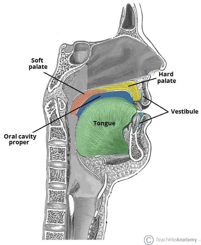
The Oral Cavity Divisions Innervation Teachmeanatomy
Anatomy Head And Neck Oral Cavity Mouth Article

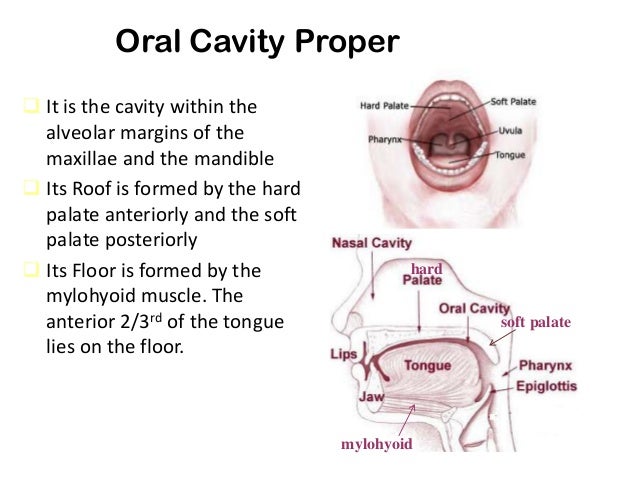
Anatomy Of The Oral Cavity Proper

Imaging The Floor Of The Mouth And The Sublingual Space Radiographics
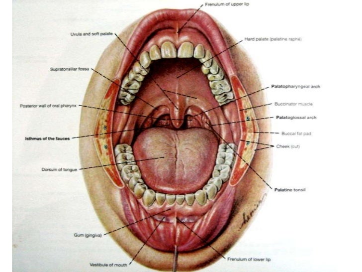
The Oral Cavity
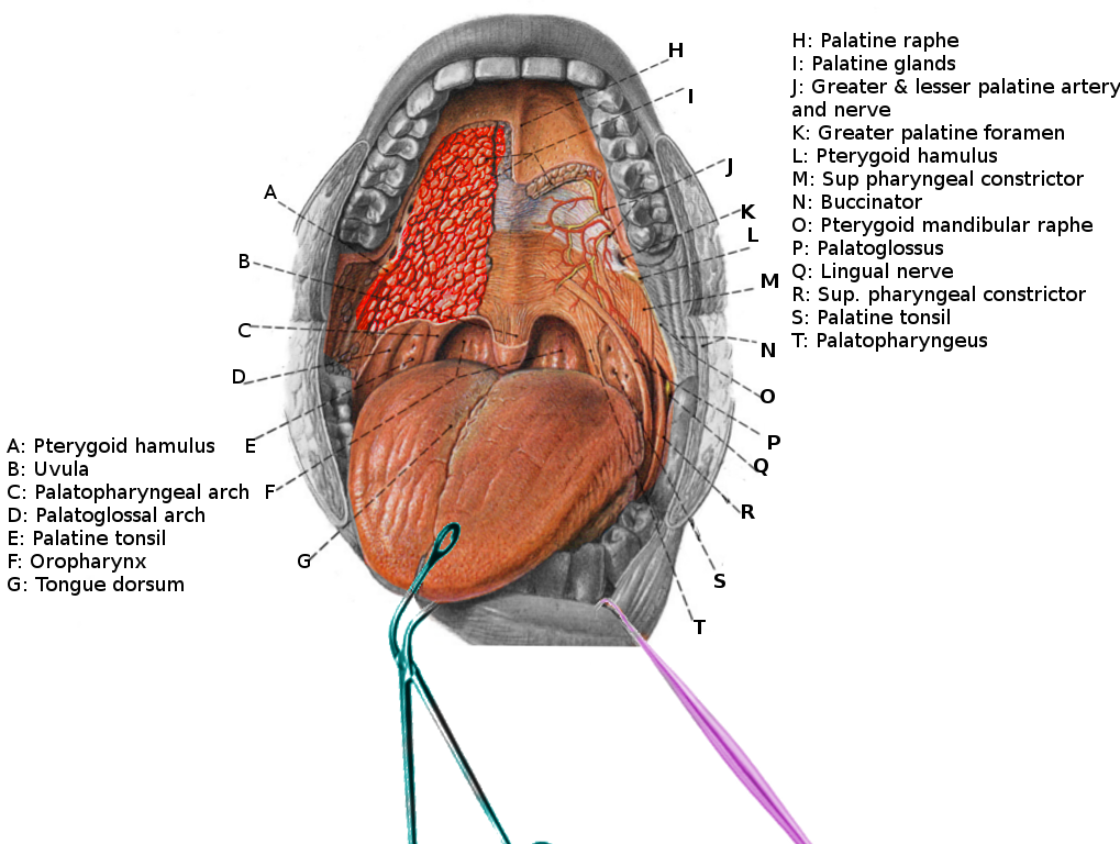
Anatomy Of Oral Region And Pharynx
Oral Cavity The Lips The Lips Are Two Fleshy Folds That Surround The Oral Orifice They Are Covered On The Outside By Skin And Are Lined On The Inside Ppt Download
Anatomy Of Oral Cavity And Oropharynx
Definition Of Oral Cavity Nci Dictionary Of Cancer Terms National Cancer Institute

Salivary Gland Anatomy Pocket Dentistry